

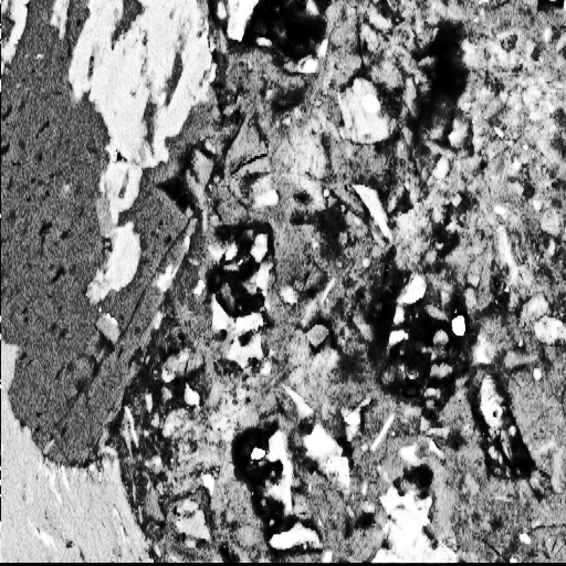
The aggregate/paste/ITZ is examined using back-scattered electron (bse) images of polished sections of concrete. Such Scanning Electron Microscope (SEM) images are subject to significant speckle. Fig. 1 shows an example, which is a 512x512 cutout of a larger 1024x1024 image, here shown histogram-equalized.
We see two ways to put the procedure of Basheer et al. (1999) on a more effective footing: firstly, define the thresholds based on distribution mixture modeling of the image greyscale intensity distribution; and, secondly, since this cannot be feasibly done on the available speckled images, first remove this speckle.
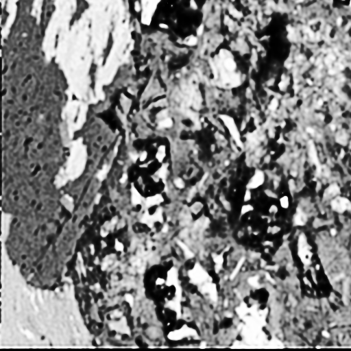
Speckle is multiplicative noise, and is typical of synthetic aperture radar (SAR) or acoustic images. The statistical modeling of speckle is discussed in Section 5.3 of MR (1999). We used the MR/1 package (MR, 1999) to despeckle the image shown in Fig. 1. The result is shown in Fig. 2 for the same 512x512 area. Despeckling the 1024x1024 image takes a few seconds elapsed time on a Sun UltraSparc 10. A wavelet transform is used, with noise modeling, to allow for speckle filtering on a range of resolution scales.
To compare the input and despeckled output images, we show a 64x64 subregion here side by side. As before, these images are shown histogram-equalized to improve the contrast.
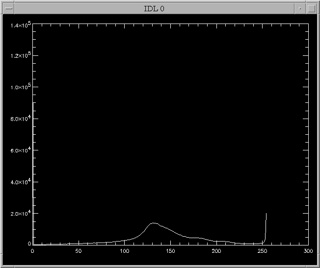
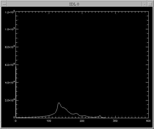
The difference in intensity distribution in these images is quite significant. Fig. 3 shows the intensity distribution of the given SEM image. It is clear than there is little by way of heuristic pointers towards where one should place thresholds leading to image segmentation. Fig. 4 shows the intensity distribution, again based on the 1024x1024 image, following filtering to remove speckle. Here, the distribution is much more amenable to the fitting of Gaussians.
We may comfortably hypothesize that, by the law of large numbers, the different constituents of the image each follow a Gaussian distribution. It is our task to disentangle these, in most cases partially, superimposed Gaussians. This can be done through an iterative fitting procedure (Celeux and Govaert, 1995). In Campbell et al. (1999), this mixture model fitting approach was used for setting thresholds in images containing various types of production faults in textiles. Furthermore in Campbell et al. (1999), we select the best model - helping significantly in selecting the best thresholds - by use of a Bayesian model selection procedure. One appropriate measure is the BIC (Bayes Information Criterion), the theory of which was developed in Kass and Raftery (1995).
Overall, this is a very effective procedure, and since the intensity distribution, only, is being processed it is highly efficient (a second or two on a normal workstation). It is an approach which we believe will lead to a solution which is quite robust for varying lighting conditions, or compositions of the concrete being studied. It is, as we have seen, capable of providing an objective procedure for the setting of the thresholds. Finally, it requires, as noted, that the images be despeckled prior to analysis.